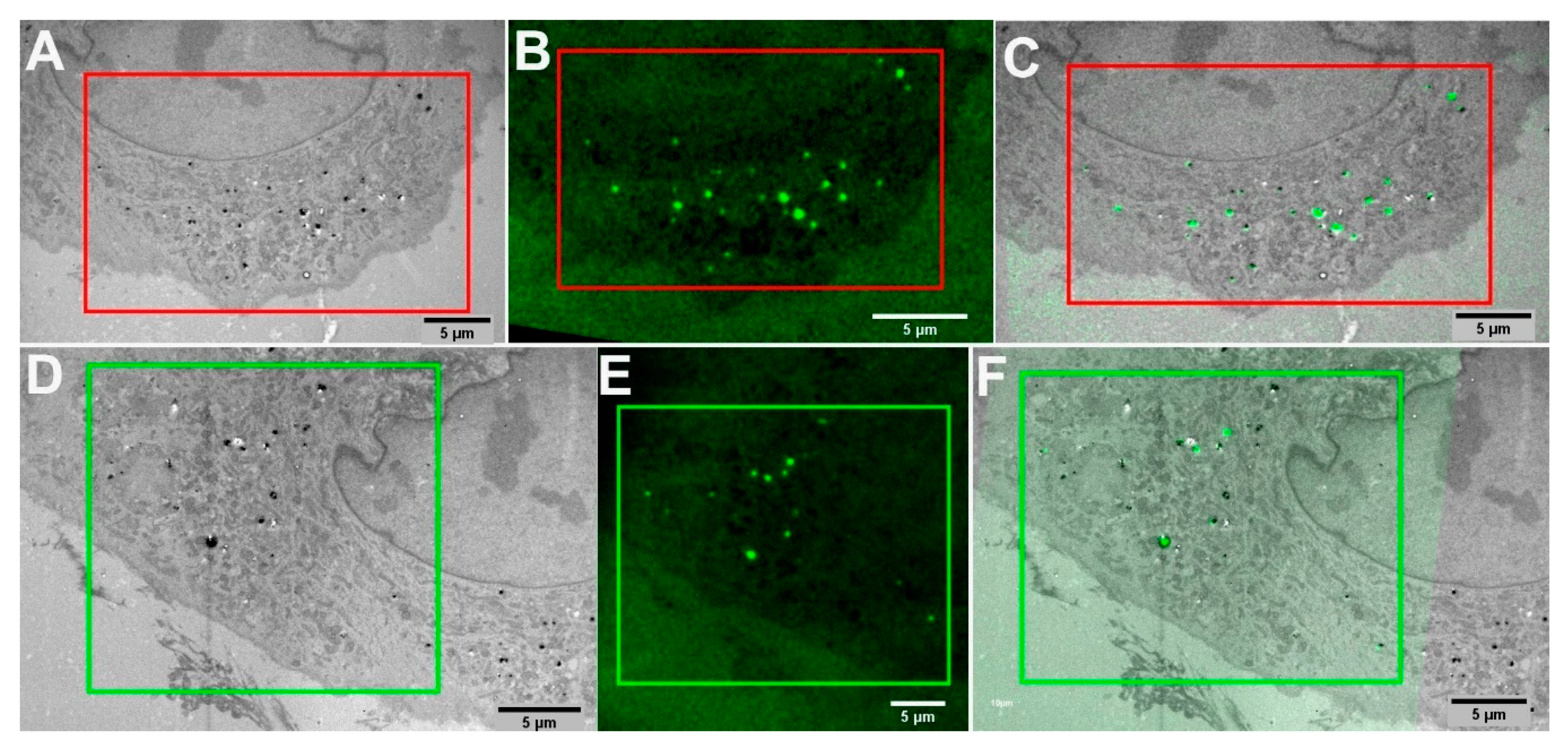
Molecules | Free Full-Text | Fluorescent and Electron-Dense Green Color Emitting Nanodiamonds for Single-Cell Correlative Microscopy
![PDF] A 25 micron-thin microscope for imaging upconverting nanoparticles with NIR-I and NIR-II illumination | Semantic Scholar PDF] A 25 micron-thin microscope for imaging upconverting nanoparticles with NIR-I and NIR-II illumination | Semantic Scholar](https://d3i71xaburhd42.cloudfront.net/58b509cad7ea5b092e45896b1818bd9d9194ad86/5-Figure1-1.png)
PDF] A 25 micron-thin microscope for imaging upconverting nanoparticles with NIR-I and NIR-II illumination | Semantic Scholar

a) SEM of the reference sample: 20 microns diameter gold FZP with 100... | Download Scientific Diagram

a) Light microscope cross-section of a spicule sample (scale bar 25... | Download Scientific Diagram
Theranostics A 25 micron-thin microscope for imaging upconverting nanoparticles with NIR-I and NIR-II illumination
![PDF] A 25 micron-thin microscope for imaging upconverting nanoparticles with NIR-I and NIR-II illumination | Semantic Scholar PDF] A 25 micron-thin microscope for imaging upconverting nanoparticles with NIR-I and NIR-II illumination | Semantic Scholar](https://d3i71xaburhd42.cloudfront.net/58b509cad7ea5b092e45896b1818bd9d9194ad86/9-Figure5-1.png)
PDF] A 25 micron-thin microscope for imaging upconverting nanoparticles with NIR-I and NIR-II illumination | Semantic Scholar
![PDF] A 25 micron-thin microscope for imaging upconverting nanoparticles with NIR-I and NIR-II illumination | Semantic Scholar PDF] A 25 micron-thin microscope for imaging upconverting nanoparticles with NIR-I and NIR-II illumination | Semantic Scholar](https://d3i71xaburhd42.cloudfront.net/58b509cad7ea5b092e45896b1818bd9d9194ad86/10-Figure6-1.png)
PDF] A 25 micron-thin microscope for imaging upconverting nanoparticles with NIR-I and NIR-II illumination | Semantic Scholar

Electron Microscope Image of 25-micron Salt Crystal Cubes « Adafruit Industries – Makers, hackers, artists, designers and engineers!
![PDF] A 25 micron-thin microscope for imaging upconverting nanoparticles with NIR-I and NIR-II illumination | Semantic Scholar PDF] A 25 micron-thin microscope for imaging upconverting nanoparticles with NIR-I and NIR-II illumination | Semantic Scholar](https://d3i71xaburhd42.cloudfront.net/58b509cad7ea5b092e45896b1818bd9d9194ad86/7-Figure4-1.png)
PDF] A 25 micron-thin microscope for imaging upconverting nanoparticles with NIR-I and NIR-II illumination | Semantic Scholar
![PDF] A 25 micron-thin microscope for imaging upconverting nanoparticles with NIR-I and NIR-II illumination | Semantic Scholar PDF] A 25 micron-thin microscope for imaging upconverting nanoparticles with NIR-I and NIR-II illumination | Semantic Scholar](https://d3i71xaburhd42.cloudfront.net/58b509cad7ea5b092e45896b1818bd9d9194ad86/6-Figure2-1.png)
PDF] A 25 micron-thin microscope for imaging upconverting nanoparticles with NIR-I and NIR-II illumination | Semantic Scholar

Optical microscope images (the scale bar denotes 200 μm length) of the... | Download Scientific Diagram

Morphology of FAAM and FDAAM. (a) Gross view, under light microscope... | Download Scientific Diagram

Greg Bright on Twitter: "Lake water under a microscope is a beautiful world most never get to see. https://t.co/MTBh1MJ81m" / Twitter
![A 25 micron-thin microscope for imaging upconverting nanoparticles with NIR-I and NIR-II illumination [Abstract] A 25 micron-thin microscope for imaging upconverting nanoparticles with NIR-I and NIR-II illumination [Abstract]](https://www.thno.org/v09/p8239/toc.jpg)


